Spina bifida
Operation part 1
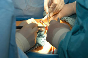
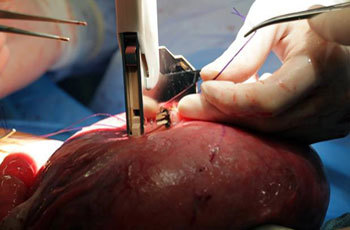
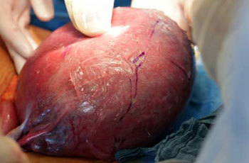
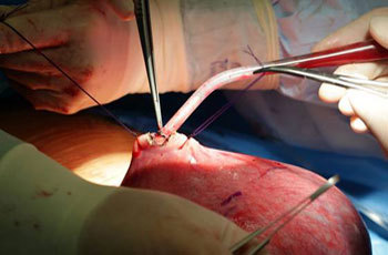
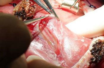
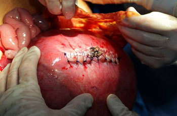
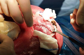
The mother’s abdomen is opened in more or less the same way as in a cesarean section (1). The uterus is lifted partially out of the abdomen (2) and the site at which it is to be opened is determined with the aid of the ultrasound transducer. A small initial opening is made in the uterine wall between holding sutures to permit the introduction of an instrument (stapler) for making a longer incision (3). A stapler is used to make a longer opening in the uterine wall, all layers of the uterine wall being clamped toigether and any bleeding stopped (4). The lesion, with its soft and histologically intact spinal cord (arrow), has been brought to the middle of the opening in the uterine wall for the operation (5).
Operation part 2
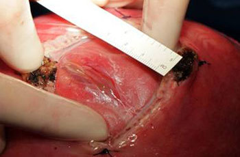
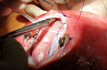
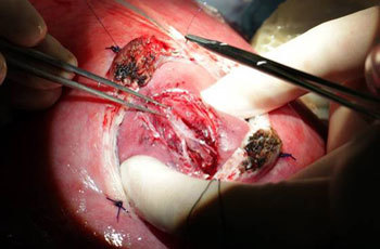
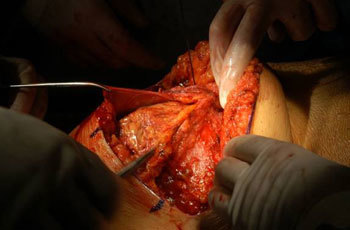
As a first step, the parts of the lesion that are to be removed are dissected free and snipped off (1). Layers of protective tissue are sewn over the spinal cord (2). In the next stage the skin on the fetus’s back is closed over the lesion to form a protective outer layer (3). The uterus is then closed with several rows of sutures to ensure a watertight seal (4). The fatty apron in the mother’s abdominal cavity is sewn over the uterine suture to form an additional sealing layer (5). The final step in the operation is to close the mother’s abdomen (6).


